Thursday 7 November 2013

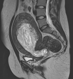
Endometrial Hyperplasia - MRI

Axial T1

Axial T2

Coronal T2


Sagittal T2

Diffuse hyperintense signal uniform heterogenous thickening of the endometrium on the T2 images that is intermediate signal on T1.

The hyperplasia extends to into the cervical canal bulging through the external os.

This is was histologically proven to be Endometrial hyperplasia.


Monday 5 August 2013

Choledochal cyst Type IV - MRI features


Coronal HASTE


Axial T1


Axial T2
             3D MIP                                    


3D MIP

38 year old female patient who presented with abdominal pain and had an ultrasound which showed dilatation of the CBD and IHRD.

MRCP showed cystic dilatation of the intrahepatic biliary ducts (arrow) with cystic dilatation of the common hepatic and bile duct (*) upto the lower third.

Diagnosis - Choledochal cyst - Type IVA


Reference:


Thursday 1 August 2013

Calcaneonavicular Coalition - Fibrous/Cartilaginous

Axial IR

Axial T1

Sagittal IR

Sagittal T1


12 year old girl presented with foot pain.

MRI demonstrated enlongated broad anterior process of the calcaneum - anteater's nose - that showed abnormal articulation with the navicular bone.

Hyperintense signal noted at the abnormal articular ends on the IR images.

Diagnosis: Calcaneonavicular coalition Fibrous/Cartilaginous type.

Most common subtype of tarsal coalition.

Reference and further reading:

http://www.radsource.us/clinic/0812


Saturday 29 June 2013

Cowper's duct cysts

T1 sagittal

T2 axial

T2 coronal

T2 sagittal

30 year old presented with dysuria and occassional hematuria.

MRI of the pelvis demonstrated:

- a well defined hyperintense cystic lesion on T1 and T2
- Lesion lies in the midline in close approximation to the bulbous urethra and just below the prostate.


Diagnosis: Cowper's duct cyst.

Cowper's duct cyst:


  • Cowpers glands - found in the urogenital diaphragm below the prostate
  • Drains into the bulbar urethra
  • Obstruction results in retention cysts
  • Cowper's gland cysts are probably secondary to trauma or infection
  • Large cysts can cause urinary obstruction, hematuria or infertility.
  • Treated by marsupialisation or endoscopic incision.
References:


Tuesday 25 June 2013

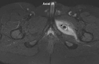
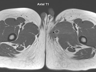
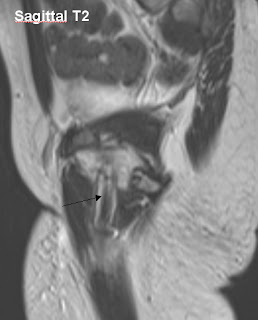
Perineal injury - Foreign body in the obturator externus - Ouch!!!


12 year old presented with a focal wound in the left aspect of the perineum.

According to the mother the boy came back from school with a perineal injury with no clear history or cause -said to be a fall. The boy initilally appeared hesitant to reveal the exact cause.

A few days later mother noticed fluid oozing from the wound with pieces of lead being extruded.

Boy was seen by a surgeon who referred him for an MRI.


Images demonstrates a linear hypointense signal structure (arrow) lying in the left obturator externus with surrounding muscle edema.

Further history according to the boy then revealed that at school an upright pencil was placed on the seat of the boy's chair by the adjacent student  as a prank just prior to the boy attempting to sit down - thus pierced through the left perineum - Ouch!!!


Friday 21 June 2013

Osteopoikilosis - MRI features

Coronal IR

Coronal T1

Coronal T1

Rorschach


Young patient who presented with low back ache and came in for an MRI of the lumbar spine.

Lumbar spine images demonstrated a marked posterior disc extrusion at L4/5 accounting for the patient’s symptoms.

However on the routine coronal IR images multiple well defined ovoid hypointense foci were noticed in the pelvic girdle and femur.

An additional coronal T1 was acquired which demonstrates multiple numerous dark foci representing bone islands

Reminded me of the facial features of Rorshach from the cult graphic novel “The  Watchmen”

Diagnosis: Osteopoikilosis

Osteopoikilosis:  

- is a sclerosing bone dysplasia with multiple 
        enostoses
- develop during childhood and do not regress and 
        therefore are seen in all age groups
- asymptomatic 
- often found concurrently with osteopathia striata, 
        and melorheostosis 
- bone islands of osteopoikilosis are typically 
        clustered around joints and align themselves        
        parallel to surrounding trabeculae
- found in the appendicular skeleton and pelvis
- axial skelton is largely spared. It is rare for the 
        skull vault to be involved 
- is one of the skeletal “Don’t touch” lesions.

References: