Pages

Saturday, 30 June 2012

MRI in neck imaging This is a power point presentation about MRI in Neck.Initial few slides are about anatomy of neck.Later cases.Cases are about MR sialogram , carotid body tumor , infiltrating nasopharyngeal carcinoma , vocal cord palsy , neurogenic tumor and CT versus MRI in neck imaging. 


Contributed by Dr Krishna Kiran

Wednesday, 20 June 2012

Large Sessile Osteochondroma Of The Lesser Trochanter Of Femur - MRI features


Osteochondroma 

- most common bone tumor
- developmental lesion rather than a true neoplasm. 
- 20%–50% of all benign bone tumors and 10%–15% of all bone tumors.
- composed of cortical and medullary bone with an overlying hyaline cartilage cap and must demonstrate continuity with the underlying parent bone cortex and medullary canal. 
- Complications  - deformity, fracture, neurovascular compromise, overlying bursa formation, and malignant transformation
- Malignant transformation is seen in 1% of solitary osteochondromas. Continued lesion growth and a hyaline cartilage cap greater than 1.5 cm in thickness, after skeletal maturity, suggest malignant transformation. 

MRI of the osteochondroma (grey solid arrow) is the best radiologic modality for visualizing the effect of the lesion on surrounding structures and evaluating the hyaline cartilage cap. 

High water content in nonmineralized portions of the cartilage cap shows intermediate to low signal intensity on T1-weighted images and very high signal intensity on T2-weighted MR images ( yellow pentagon).


MRI allows accurate measurement of the cartilage cap thickness and distinction from overlying muscle on MR images. 

References:

 Murphey MD et al :Imaging of Osteochondroma: Variants and Complications with Radiologic-Pathologic Correlation - Radiographics September 2000 20:1407-1434


BASICS OF MRI

Contributed by Dr Krishna Kiran

Azgos Anterior cerebral artery


Azygos anterior cerebral artery


An azygos anterior cerebral artery is an uncommon to rare variant seen in approximately 0.4 -1% of the population  in which the two A1 segments of the ACA join to form a single trunk. As a result no anterior communicating artery is present. This organization is similar to that seen in lower primates .
It is associated with numerous abnormalities, including:
dysgenesis of the corpus callosum
lobar holoprosencephaly 
septooptic dysplasia
porencephalic cysts
arteriovenous malformations (AVM)
Berry aneurysms are often seen at the eventual bifurcation of the single vessel, due to either altered haemodynamics or congenitally abnormal wall or both. The reported incidence varies widely from 13 to 71% .
As there is frequent a) hypoplasia of the ACOM and b) contralateral supply of the medial surface of the hemispheres by the ACA branches, confirmation of an azygos ACA can be difficult on angiography . Cross compression views can be helpful.

References

http://radiopaedia.org/articles/azygos-anterior-cerebral-artery

Friday, 15 June 2012

Colo-colic intussusception on MR enterography

Coronal

MR enterography was performed on a 30 year old patient who had complained about intermittent abdominal pain.


Colo- colic intussusception was detected with the cecal pole appearing to be the leading point (intussusceptum) and pulling up the ileocaecal junction along with past the hepatic flexure


This was secondary to a caecal adenocarcinoma contributing to the intussusceptum.

Tuesday, 12 June 2012

Iliotibial Band syndrome - Hip


                                                                          Axial STIR


                                                                         Coronal STIR


                                                                             Axial T1


Patient presenting with left lateral hip pain.


Diffuse thickening of the proximal left iliotibial band is seen with surrounding hyperintensity on  the STIR images


Iliotibial band syndrome is not an uncommon source of knee and hip pain  especially in running athletes.


Irritation and inflammation can occur as the band rubs across , commonly the lateral femoral condyle and less commonly the greater trochanter.


Testicular pain in a young male


24 years young male complains of severe pain in right testis since two days.
USG shows hypoechoic lesion near lower pole. MRI was done.


what do you think is the diagnosis..?

Let us see the findings first.

arrow points to a triangular hypointensity.


arrows point to a triangular hypointensity.


an area of facilitated diffusion seen in lower pole of right testis.


T1 contrast and in particular subtraction image shows area of lack of enhancement surrounded by well enhancing rim.


These findings with history of pain are consistent with segmental testicular infarct.

what are radiological findings of segmental testicular infarction.
It is characterised by presence of a triangular shaped avascular intrataesticular lesion on sonography or MRI and enhancement of surrounding borders on MRI.

how do you differentiate it from testicular tumor.
triangular shape , lack of vascularity , clinical setting of severe pain are differenital features of segmental infarct. follow up should be advised.

what is the follow up in this patient.
this patient is under observation and is getting better.

reference
Gabriel Fernandez et al. radiologic findings of segmental testicular infarction.AJR May 2005


Monday, 11 June 2012

MR Urography of Sober Ureterostomy


Follow up MR urography in a patient who had undergone bilateral Sober ureterostomy for posterior urethral valve.

This procedure involves the division of the ureter with reanastomosis of the distal limb to the side of the proximal limb (arrows) . The proximal limb extends to the cutaneous surfaces covered by ureterostomy bags.

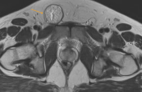
Coracoclavicular joint


This patient presented with pain  in the region of the lateral aspect of the left clavicle.


Congenital costoclavicular joint is rare and is usually a bilateral anatomic variant and asymptomatic.

In the absence of other pathology in cases of shoulder pain these joints may produce symptoms of supracoracoid impingement ( pain on flexion and neutral rotation).

Wednesday, 6 June 2012

Right Diaphragmatic Hernia


A case of a 14 year old boy who had a history of previous congenital hernia repair.


Presented with recurrent diaphragmatic hernia.


Note the herniation of the liver, hepatic flexure and loops of small bowel ( grey solid arrow) into the right lower lung-thus collapsing the right lower lobe .


On the sagittal images note the stretched mesentery and mesenteric vessels across the anterior surface of the liver (yellow pentagon).


Diagnosis: Recurrent diaphragmatic hernia


Radial Meniscal tear


22 year old man with a history of injury 2 months ago.


There is a high signal defect in the inner margin of the posterior horn of the medial meniscus on the coronal PD and PDFS sequences ( yellow pentagon).


On the sagittal images the defect is seen as a "truncated triangle" ( grey solid arrow).


Appearances are indicative of a radial tear 

Saturday, 2 June 2012

Ovarian Torsion


(a) Coronal T2 


                                                               (b) Sagittal T2 


                                                               (c) Axial T2 - fat sat 


                                                                   (d) Axial T1 


                                     (e) Coronal T2 


18 year old girl presented with left sided pelvic pain.


Coronal & Axial T2 images demonstrate an enlarged engorged congested left ovary (grey arrow) that demonstrates a hyperintense central stroma with peripheral follicles 


There is a large thick-walled right sided exophytic ovarian cyst (yellow pentagon) seen on the (c)T2 fat sat images.


There is associated free fluid in the pelvis (curved arrow).


The T1 images (d) also shows a mildly hyperintense stroma indicating congestion.


On the magnified coronal T2 (e) note the twisted fallopian tube  (notched arrow).


In this case the uterus was noted to be pulled towards the side of the torted ovary.


Conclusion: Enlarged congested left ovary with coexistent cyst and twisted pedicle is pathognomonic of ovarian torsion.