Pages

Monday, 30 July 2012

Isolated hypoglossal nerve palsy due to a tortuous vertebral artery course -MRI features

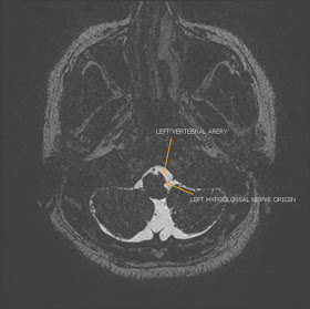
42 year old man presenting with difficulty in speech and deviation of the tongue to the left. MRI was performed to assess for organic cause.


Axial 3D SPACE


Axial T1


Coronal STIR


MRI showed a demonstrated a tortuous left vertebral artery compressing the medulla at the emergence of the left hypoglossal nerve rootlets.


On the coronal STIR images there is atrophy of the left half of  the tongue which appears reduced in bulk and minimally hyperintense compared to the right.


Microvascular decompression of the vertebral artery can result in resolution of the nerve palsy. 


Monday, 16 July 2012

Os acromiale - mesoacromion -symptomatic - MRI features


27 year old female patient with left shoulder pain. No history of trauma.


Coronal Oblique PDFS

Axial T1


MRI shows presence of os acromiale with hyperintense signal seen at the synchondrosis on the PDFS images indicative of edema which could be the cause of the pain.

There was no evidence of secondary rotator cuff impingement.

MRI shows presence of os acromiale with hyperintense signal seen at the synchondrosis on the STIR images indicative of edema which could be the cause of the pain.


Os acromiale:


results from failure of an anterior acromial ossification center to fuse to the  acromial process
caused by failure of one of three ossification centers; 
ossification centers appear at age 15 and should fuse by age 22-25. 
subtypes: (from anterior to posterior)
             -  preacromion, mesacromion, metacromion, and basiacromion types; 
              - mesacromion and metacromion are most common; 
patients may show impingement like symptoms but will also have 
           localized tenderness.


Tuesday, 3 July 2012

Fibula Stress Fracture - MRI features

25 year old badmintom player complaining of pain along the lateral aspect of the left leg:


Axial STIR


Axial T2


Coronal T1

Sagittal T1


Diffuse periosteal thickening is seen along the lateral cortex of the distal fibular shaft with hyperintense signal on T2 and STIR images (arrows) and hyperintense signal of the adjacent intramedullary marrow.

In the midst of the marrow hyperintensity there is a subtle horizontal linear low signal seen best on coronal T1 and sagittal T1 images (arrow) indicative of a fracture line.

There is associated surrounding soft tissue edema.


Appearances  represent a distal fibular stress fracture .

Fibular fractures account for 10% of the stress fractures and are commonly seen in the distal two thirds.