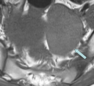
(a) Coronal T2
(b) Sagittal T2
(c) Axial T2 - fat sat
(d) Axial T1
(e) Coronal T2
18 year old girl presented with left sided pelvic pain.
Coronal & Axial T2 images demonstrate an enlarged engorged congested left ovary (grey arrow) that demonstrates a hyperintense central stroma with peripheral follicles
There is a large thick-walled right sided exophytic ovarian cyst (yellow pentagon) seen on the (c)T2 fat sat images.
There is associated free fluid in the pelvis (curved arrow).
The T1 images (d) also shows a mildly hyperintense stroma indicating congestion.
On the magnified coronal T2 (e) note the twisted fallopian tube (notched arrow).
In this case the uterus was noted to be pulled towards the side of the torted ovary.
Conclusion: Enlarged congested left ovary with coexistent cyst and twisted pedicle is pathognomonic of ovarian torsion.


No comments:
Post a Comment