


 |
Coronal HASTE |
 |
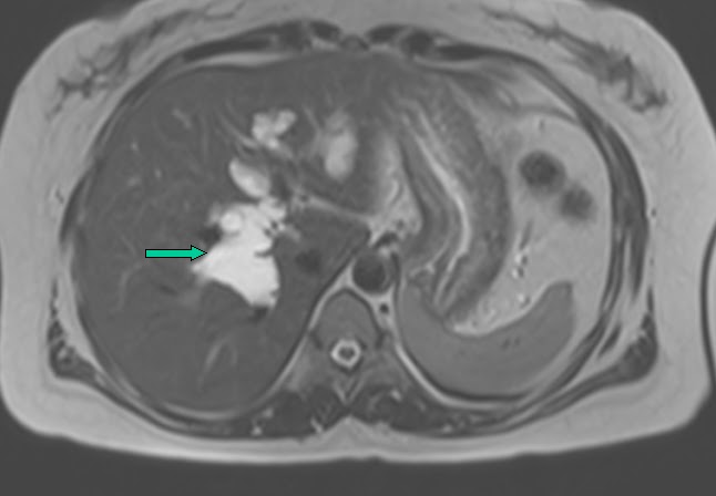 |
| Axial T2 |
 |
| 3D MIP |
 |
3D MIP 38 year old female patient who presented with abdominal pain and had an ultrasound which showed dilatation of the CBD and IHRD.
MRCP showed cystic dilatation of the intrahepatic biliary ducts (arrow) with cystic dilatation of the common hepatic and bile duct (*) upto the lower third.
Diagnosis - Choledochal cyst - Type IVA

Reference:
|
 |
| Axial IR |
 |
| Axial T1 |
 |
| Sagittal IR |
 |
| Sagittal T1
12 year old girl presented with foot pain.
MRI demonstrated enlongated broad anterior process of the calcaneum - anteater's nose - that showed abnormal articulation with the navicular bone.
Hyperintense signal noted at the abnormal articular ends on the IR images.
Diagnosis: Calcaneonavicular coalition Fibrous/Cartilaginous type.
Most common subtype of tarsal coalition.
Reference and further reading:
http://www.radsource.us/clinic/0812
|