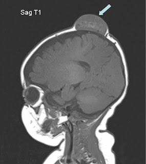
3 month old with scalp swelling
Well delineated subcutaneous scalp mass ( block arrow) in the left parasagittal frontoparietal region that shows intermediate to high signal on T1 and T2 images with linear flow voids (small arrow heads). No intracranial extension.
MRI is usually performed to characterize the swelling and to exclude intracranial extension.


No comments:
Post a Comment